- Electronic Bonus Plates
- General Anatomy
- Surface Anatomy
- Electronic Bonus Plates
صفحه
مدرسه پزشکی

Netter Atlas of Human Anatomy: Classic Regional Approach
For students and clinical professionals who are learning anatomy, participating in a dissection lab, sharing anatomy knowledge with patients, or refreshing their anatomy knowledge, the Netter Atlas of Human Anatomy illustrates the body, region by region, in clear, brilliant detail from a clinician’s perspective. Unique among anatomy atlases, it contains illustrations that emphasize anatomic relationships that are most important to the clinician in training and practice. Illustrated by clinicians, for clinicians, it contains more than 550 exquisite plates plus dozens of carefully selected radiologic images for common views.
درخواست و رزرو جلسه مصاحبه
ویژگی های این کتاب

World-Renowned Medical Illustrations
شامل آثار هنری آناتومیک نقاشیشده با دست اثر نمادین فرانک اچ. نتر با وضوح و دقت استثنایی.
Comprehensive Regional Anatomy
بدن انسان را بر اساس نواحی آناتومیک برای یادگیری منطقی، ساختارمند و کارآمد سازماندهی میکند.
Clinically Relevant Visualization
تصاویر آناتومیک دقیق را با عملکرد بالینی دنیای واقعی و درک تشخیصی مرتبط میکند.
The World's Most Trusted Anatomy Atlas
اطلس آناتومیک مورد علاقه برای دانشجویان پزشکی، مربیان، جراحان و متخصصان مراقبتهای بهداشتی در سراسر جهان.
Contents
- Electronic Bonus Plates
- Overview
- Spinal Cord
- Brain
- Cerebral Vasculature
- Cranial and Cervical Nerves
- Eye
- Nerves of Thorax
- Electronic Bonus Plates
- Overview
- Cranium, Mandible, and Temporomandibular Joint
- Vertebral Column
- Thoracic Skeleton
- Bony Pelvis
- Upper Limb
- Lower Limb
- Electronic Bonus Plates
- Overview
- Head and Neck
- Back
- Thorax
- Abdomen and Pelvis
- Upper Limb
- Lower Limb
- Structures with High Clinical Significance
- Electronic Bonus Plates
- Electronic Bonus Plates
- Pericardium
- Heart
- Blood Vessels of the Head and Neck
- Blood Vessels of the Limbs
- Blood Vessels of the Trunk
- Structures with High Clinical Significance
- Electronic Bonus Plates
- Electronic Bonus Plates
- Head and Neck
- Limbs
- Trunk
- Structures with High Clinical Significance
- Electronic Bonus Plates
- Electronic Bonus Plates
- Nasal Cavity
- Vasculature and Innervation of Nasal Cavity
- Paranasal Sinuses
- Pharynx
- Larynx
- Trachea and Bronchi
- Vasculature and Innervation of Tracheobronchial Tree
- Lungs
- Vasculature and Innervation of Mediastinum
- Electronic Bonus Plates
- Mouth
- Pharynx
- Viscera (Esophagus, Stomach, Intestines, Liver, Pancreas)
- Visceral Vasculature
- Visceral Innervation
- Structures with High Clinical Significance
- Electronic Bonus Plates
- Electronic Bonus Plates
- Kidneys and Ureter
- Urinary Bladder and Urethra
- Vasculature and Innervation
- Structures with High Clinical Significance
- Electronic Bonus Plates
- Electronic Bonus Plates
- Mammary Glands
- Bony Pelvis
- Pelvic Diaphragm and Pelvic Cavity
- Female Internal Genitalia
- Female Perineum and External Genitalia
- Male Internal Genitalia
- Male Perineum and External Genitalia
- Homologies of Male and Female Genitalia
- Imaging of Pelvic Viscera
نمونه ای از آنچه خواهید آموخت
Skin
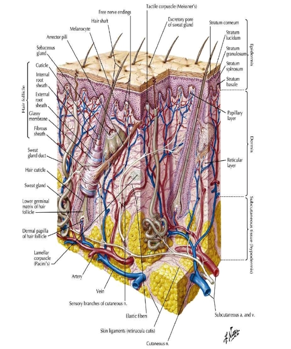
The human skin is the outer covering of the body and is the largest organ of the integumentary system. The skin has up to seven layers of ectodermal tissue guarding muscles, bones, ligaments and internal organs. Human skin is similar to most of the other mammals' skin, and it is very similar to pig skin. Though nearly all human skin is covered with hair follicles, it can appear hairless. There are two general types of skin: hairy and glabrous skin (hairless). The adjective cutaneous literally means "of the skin" (from Latin cutis, skin).Skin plays an important immunity role in protecting the body against pathogens and excessive water loss. Its other functions are insulation, temperature regulation, sensation, synthesis of vitamin D, and the protection of vitamin B folates...
The central nervous system
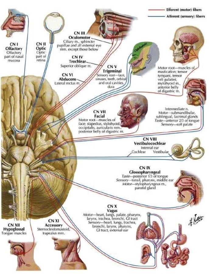
The central nervous system (CNS) is the part of the nervous system consisting primarily of the brain, and the spinal cord. The retina is also technically part of the CNS.The CNS is named so because the brain integrates the received information and coordinates and influences the activity of all parts of the bodies of bilaterally symmetric and triploblastic animals—that is, all multicellular animals except sponges and diploblasts. It is a structure composed of nervous tissue positioned along the rostral (nose end) to caudal (tail end) axis of the body and may have an enlarged section at the rostral end which is a brain. Only arthropods, cephalopods and vertebrates have a true brain, though precursor structures exist in onychophorans, gastropods and lancelets...
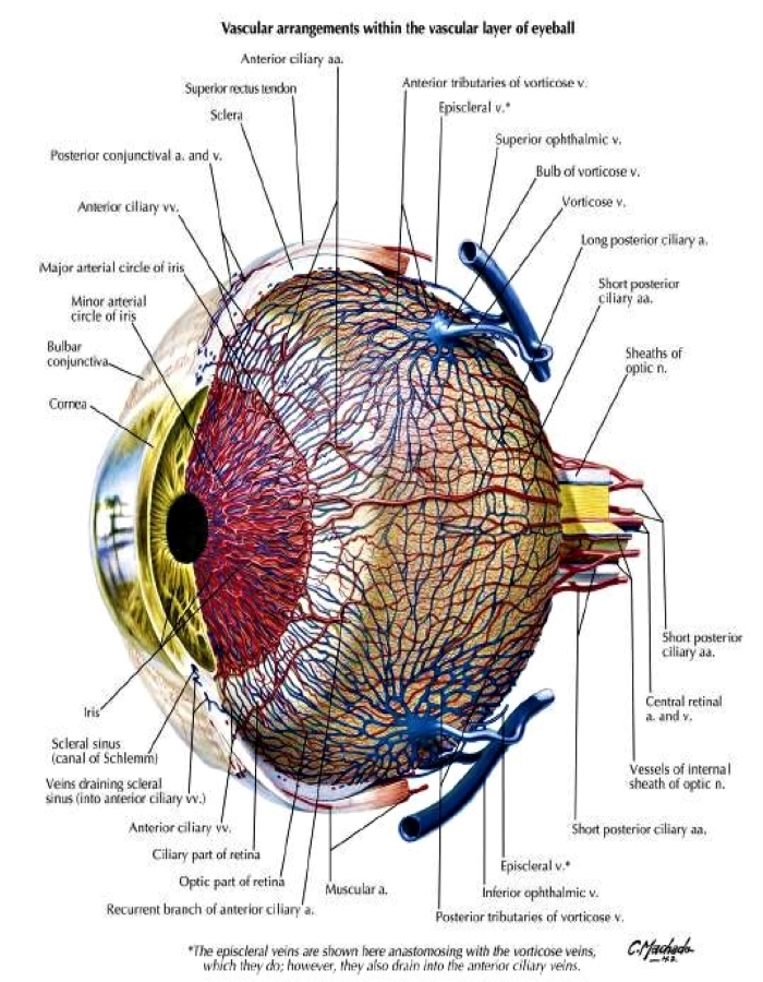
Eye
An eye is a sensory organ that allows an organism to perceive visual information. It detects light and converts it into electro-chemical impulses in neurons (neurones). It is part of an organism's visual system. In higher organisms, the eye is a complex optical system that collects light from the surrounding environment, regulates its intensity through a diaphragm, focuses it through an adjustable assembly of lenses to form an image, converts this image into a set of electrical signals, and transmits these signals to the brain through neural pathways that connect the eye via the optic nerve to the visual cortex and other areas of the brain. Eyes with resolving power have come in ten fundamentally different forms, classified into compound...
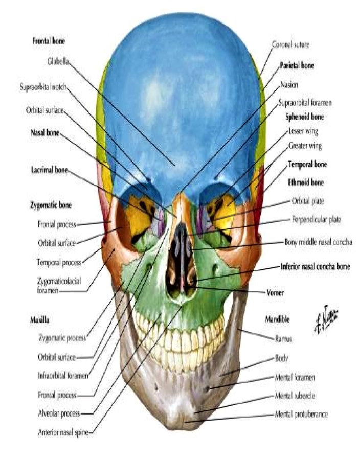
Skull
The skull, or cranium, is typically a bony enclosure around the brain of a vertebrate.In some fish and amphibians, the skull is of cartilage. The skull is at the head end of the vertebrate. In a human, the skull comprises two prominent parts: the neurocranium and the facial skeleton,which evolved from the first pharyngeal arch. The skull forms the frontmost portion of the axial skeleton and is a product of cephalization and vesicular enlargement of the brain, with several special senses structures such as the eyes, ears, nose, tongue and, in fish, specialized tactile organs such as barbels near the mouth. The skull is composed of three types of bone: cranial bones, facial bones and ossicles, which is made up of a number of fused flat and irregular bones...
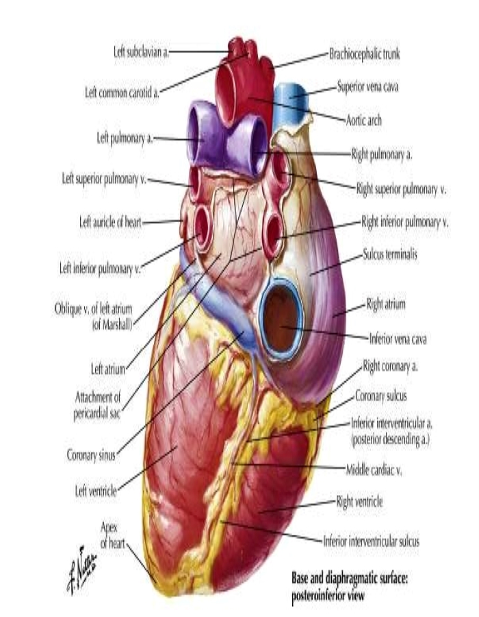
Heart
The heart is a muscular organ found in humans and other animals. This organ pumps blood through the blood vessels.The heart and blood vessels together make up the circulatory system.The pumped blood carries oxygen and nutrients to the tissue, while carrying metabolic waste such as carbon dioxide to the lungs.In humans, the heart is approximately the size of a closed fist and is located between the lungs, in the middle compartment of the chest, called the mediastinum. In humans, the heart is divided into four chambers: upper left and right atria and lower left and right ventricles. Commonly, the right atrium and ventricle are referred together as the right heart and their left counterparts as the left heart. In a healthy heart, blood flows one way through the heart due to heart valves, which prevent backflow...
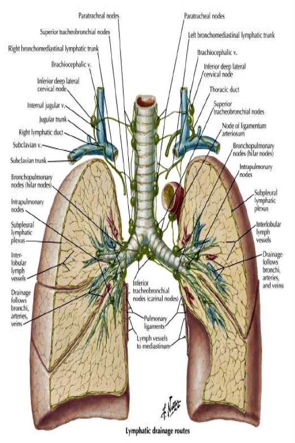
Lung
The lungs are the primary organs of the respiratory system in many animals, including humans. In mammals and most other tetrapods, two lungs are located near the backbone on either side of the heart. Their function in the respiratory system is to extract oxygen from the atmosphere and transfer it into the bloodstream, and to release carbon dioxide from the bloodstream into the atmosphere, in a process of gas exchange. Respiration is driven by different muscular systems in different species. Mammals, reptiles and birds use their musculoskeletal systems to support and foster breathing. In early tetrapods, air was driven into the lungs by the pharyngeal muscles via buccal pumping, a mechanism still seen in amphibians. In humans, the primary muscle that drives breathing is the diaphragm...
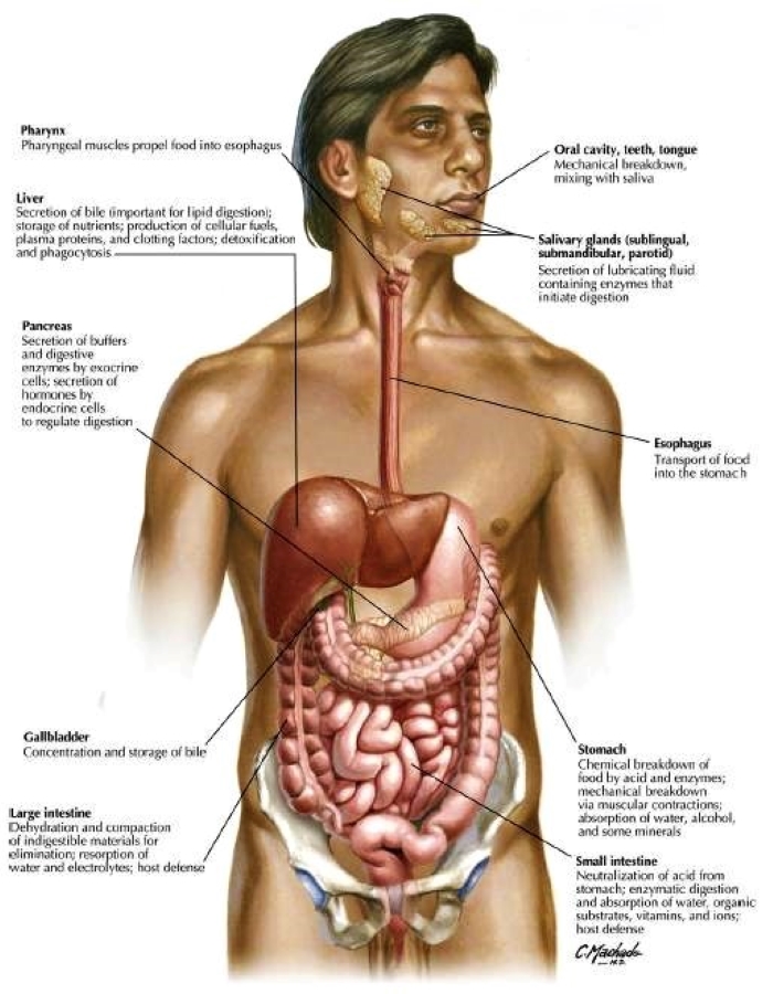
Digestive system
The human digestive system consists of the gastrointestinal tract plus the accessory organs of digestion (the tongue, salivary glands, pancreas, liver, and gallbladder). Digestion involves the breakdown of food into smaller and smaller components, until they can be absorbed and assimilated into the body. The process of digestion has three stages: the cephalic phase, the gastric phase, and the intestinal phase. The first stage, the cephalic phase of digestion, begins with secretions from gastric glands in response to the sight and smell of food, and continues in the mouth with the mechanical breakdown of food by chewing, and the chemical breakdown by digestive enzymes in the saliva. Saliva contains amylase, and lingual lipase, secreted by the salivary glands, and serous glands on the tongue...
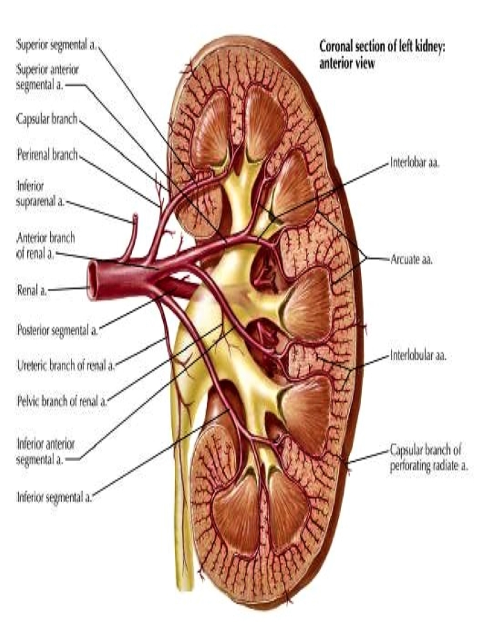
Kidney
In humans, the kidneys are two reddish-brown bean-shaped blood-filtering organs[1] that are a multilobar, multipapillary form of mammalian kidneys, usually without signs of external lobulation.[2][3] They are located on the left and right in the retroperitoneal space, and in adult humans are about 12 centimetres (4+1⁄2 inches) in length.[4][5] They receive blood from the paired renal arteries; blood exits into the paired renal veins. Each kidney is attached to a ureter, a tube that carries excreted urine to the bladder. The kidney participates in the control of the volume of various body fluids, fluid osmolality, acid–base balance, various electrolyte concentrations, and removal of toxins. Filtration occurs in the glomerulus: one-fifth of the blood volume that enters the kidneys is filtered.Examples of substances reabsorbed are solute-free water...
مدسه پزشکی در مسیر موفقیت فرزندتان

فرم درخواست رزرو مصاحبه
شروع کلاس های جدید مدرسه پزشکی در مرداد ماه سال 1405
تمرکز اصلی بر روزهای چهارشنبه، پنجشنبه و جمعه
حضوری تهران و همزمان آنلاین در سراسر کشور
36 ماه آموزش تخصصی
پشتیبانی هر روزه در طول این 36 ماه آموزش تخصصی