- Chapter 1: An Introduction to the Human Body
- Chapter 2: The Chemical Level of Organization
- Chapter 3: The Cellular Level of Organization
- Chapter 4: The Tissue Level of Organization
صفحه
مدرسه پزشکی

OpenStax: Anatomy and Physiology
Anatomy and Physiology 2e is developed to meet the scope and sequence for a two-semester human anatomy and physiology course for life science and allied health majors. The book is organized by body systems. The revision focuses on inclusive and equitable instruction and includes new student support. Illustrations have been extensively revised to be clearer and more inclusive...
درخواست و رزرو جلسه مصاحبهویژگی های این کتاب

Comprehensive Coverage
کاوش در تمام سیستمهای اصلی بدن، از ساختار سلولی گرفته تا عملکرد اندامها

Evidence-Based Content
بر اساس استانداردهای دانشگاهی شناخته شده بین المللی ساخته شده است

Clinical Relevance
دانش آناتومی را با فرآیندهای فیزیولوژیکی و کاربردهای مراقبتهای بهداشتی مرتبط کنید

Ideal for Healthcare Students
مناسب برای رشتههای پزشکی، پرستاری، فیزیوتراپی، زیستشناسی و علوم وابسته به سلامت
Contents
- Chapter 5: The Integumentary System
- Chapter 6 : Bone and Skeletal Tissue
- Chapter 7: The Axial Skeleton
- Chapter 8: The Appendicular Skeleton
- Chapter 9: Joints
- Chapter 10 : Muscle Tissue
- Chapter 11: The Muscular System
- Chapter 12 : Introduction to the Nervous System
- Chapter 13: The Anatomy of the Nervous System
- Chapter 14 : The Somatic Nervous System
- Chapter 15 : The Autonomic Nervous System
- Chapter 16 : The Neurological Exam
- Chapter 17: The Endocrine System
- Chapter 18 : Blood
- Chapter 19: The Cardiovascular System: The Heart
- Chapter 20: The Cardiovascular System: Blood Vessels and Circulation
- Chapter 21: The Lymphatic System and Immunity
- Chapter 22: The Respiratory System
- Chapter 23: The Digestive System
- Chapter 24: Nutrition and Metabolism
- Chapter 25: The Urinary System
- Chapter 26: Fluid, Electrolyte, and Acid–Base Balance
- Chapter 27 : The Reproductive System
- Chapter 28 : Development and Genetic Inheritance
نمونه ای از آنچه خواهید آموخت
Endocrine System
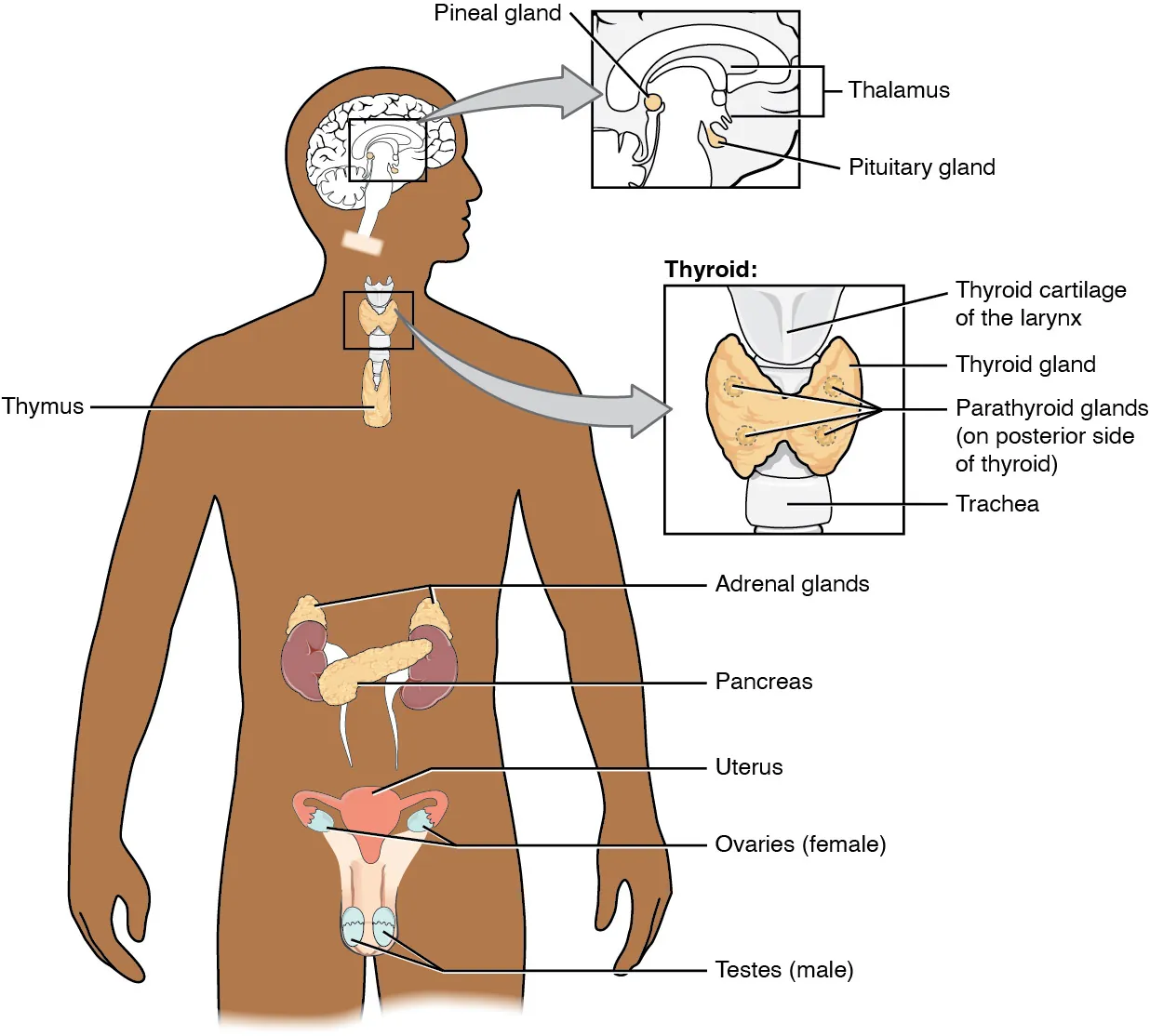
The endocrine system consists of cells, tissues, and organs that secrete hormones as a primary or secondary function. The endocrine gland is the major player in this system. The primary function of these ductless glands is to secrete their hormones directly into the surrounding fluid. The interstitial fluid and the blood vessels then transport the hormones throughout the body. The endocrine system includes the pituitary, thyroid, parathyroid, adrenal, and pineal glands (Figure 17.2). Some of these glands have both endocrine and non-endocrine functions. For example, the pancreas contains cells that function in digestion as well as cells that secrete the hormones insulin and glucagon, which regulate blood glucose levels. The hypothalamus, thymus, heart, kidneys, stomach, small intestine, liver, skin, female ovaries, and male testes are other organs that contain cells with endocrine function. Moreover, adipose tissue has long been known to produce hormones, and recent research has revealed that even bone tissue has endocrine functions...
Nervous System
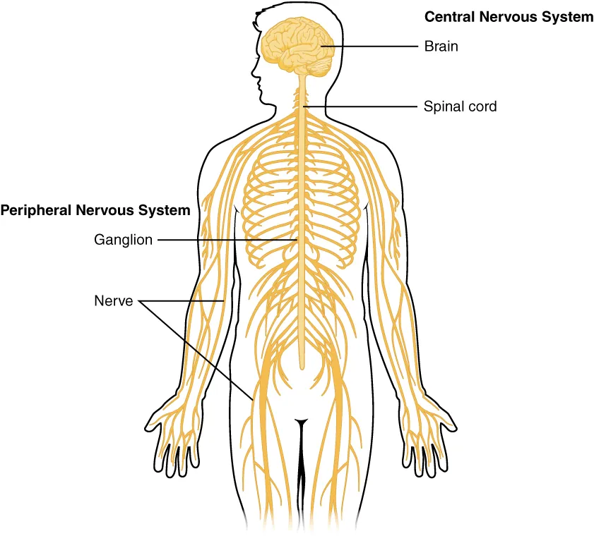
The nervous system can be divided into two major regions: the central and peripheral nervous systems. The central nervous system (CNS) is the brain and spinal cord, and the peripheral nervous system (PNS) is everything else (Figure 12.2). The brain is contained within the cranial cavity of the skull, and the spinal cord is contained within the vertebral cavity of the vertebral column. It is a bit of an oversimplification to say that the CNS is what is inside these two cavities and the peripheral nervous system is outside of them, but that is one way to start to think about it. In actuality, there are some elements of the peripheral nervous system that are within the cranial or vertebral cavities. The peripheral nervous system is so named because it is on the periphery—meaning beyond the brain and spinal cord. Depending on different aspects of the nervous system, the dividing line between central and peripheral is not necessarily universal...
Muscular System
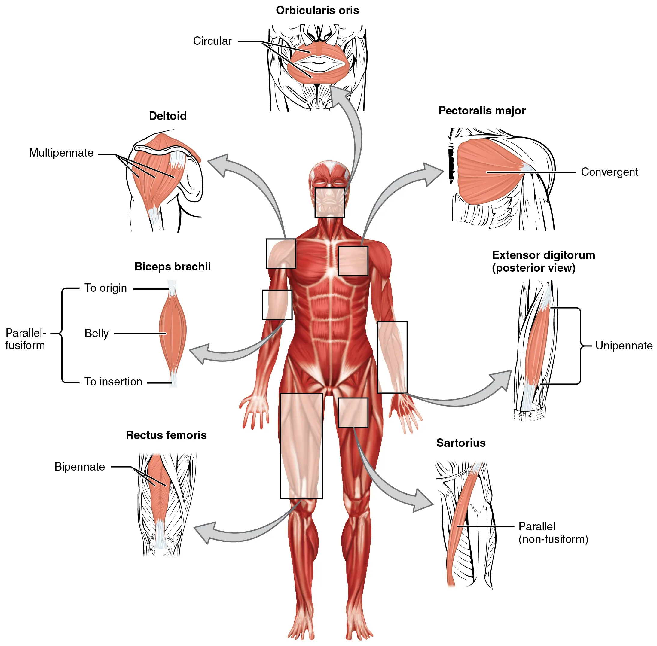
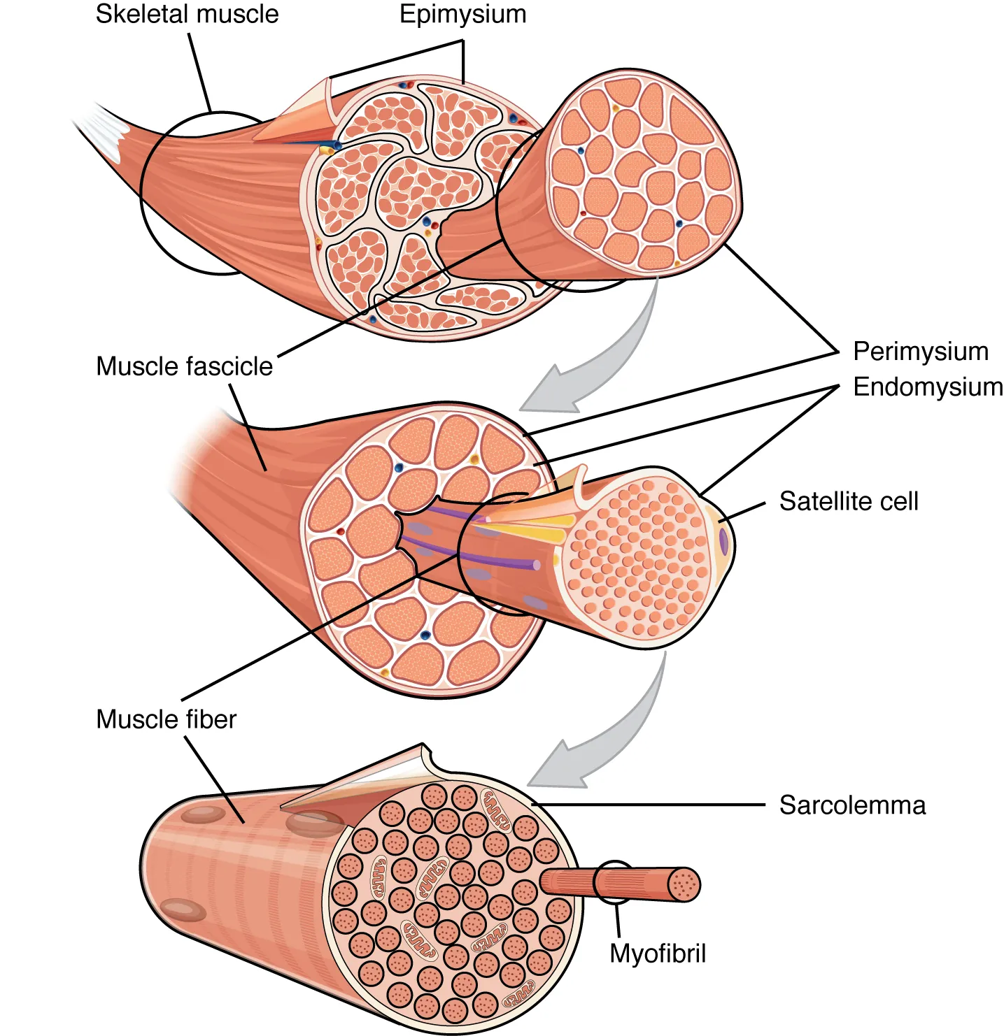
Skeletal muscle is enclosed in connective tissue scaffolding at three levels. Each muscle fiber (cell) is covered by endomysium and the entire muscle is covered by epimysium. When a group of muscle fibers is “bundled” as a unit within the whole muscle by an additional covering of a connective tissue called perimysium, that bundled group of muscle fibers is called a fascicle. Fascicle arrangement by perimysia is correlated to the force generated by a muscle; it also affects the range of motion of the muscle. Based on the patterns of fascicle arrangement, skeletal muscles can be classified in several ways. What follows are the most common fascicle arrangements. Parallel muscles have fascicles that are arranged in the same direction as the long axis of the muscle (Figure 11.3). The majority of skeletal muscles in the body have this type of organization. Some parallel muscles are flat sheets that expand at the ends to make broad attachments. Other parallel muscles are rotund with tendons at one or both ends...
Skeletal System
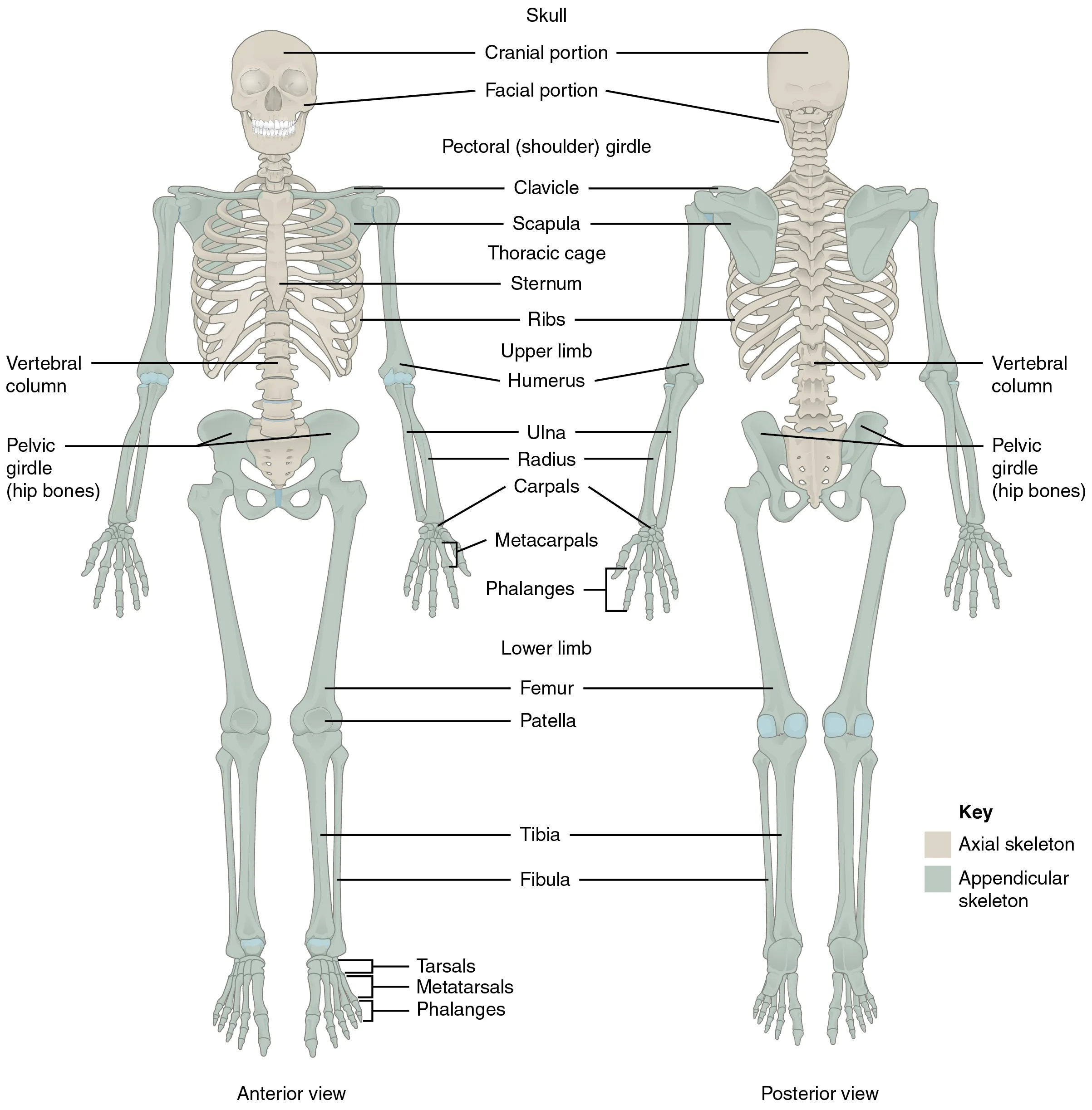
The skeletal system includes all of the bones, cartilages, and ligaments of the body that support and give shape to the body and body structures. The skeleton consists of the bones of the body. For adults, there are 206 bones in the skeleton. Younger individuals have higher numbers of bones because some bones fuse together during childhood and adolescence to form an adult bone. The primary functions of the skeleton are to provide a rigid, internal structure that can support the weight of the body against the force of gravity, and to provide a structure upon which muscles can act to produce movements of the body. The lower portion of the skeleton is specialized for stability during walking or running. In contrast, the upper skeleton has greater mobility and ranges of motion, features that allow you to lift and carry objects or turn your head and trunk. In addition to providing for support and movements of the body, the skeleton has protective and storage functions. It protects the internal organs, including the brain, spinal cord, heart, lungs, and pelvic organs...
Thyroid Gland
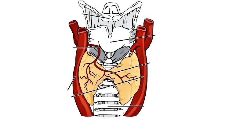
A butterfly-shaped organ, the thyroid gland is located anterior to the trachea, just inferior to the larynx (Figure 17.12). The medial region, called the isthmus, is flanked by wing-shaped left and right lobes. Each of the thyroid lobes are embedded with parathyroid glands, primarily on their posterior surfaces. The tissue of the thyroid gland is composed mostly of thyroid follicles. The follicles are made up of a central cavity filled with a sticky fluid called colloid. Surrounded by a wall of epithelial follicle cells, the colloid is the center of thyroid hormone production, and that production is dependent on the hormones’ essential and unique component: iodine.Hormones are produced in the colloid when atoms of the mineral iodine attach to a glycoprotein, called thyroglobulin, that is secreted into the colloid by the follicle cells. The following steps outline the hormones’ assembly...
Nervous Tissue
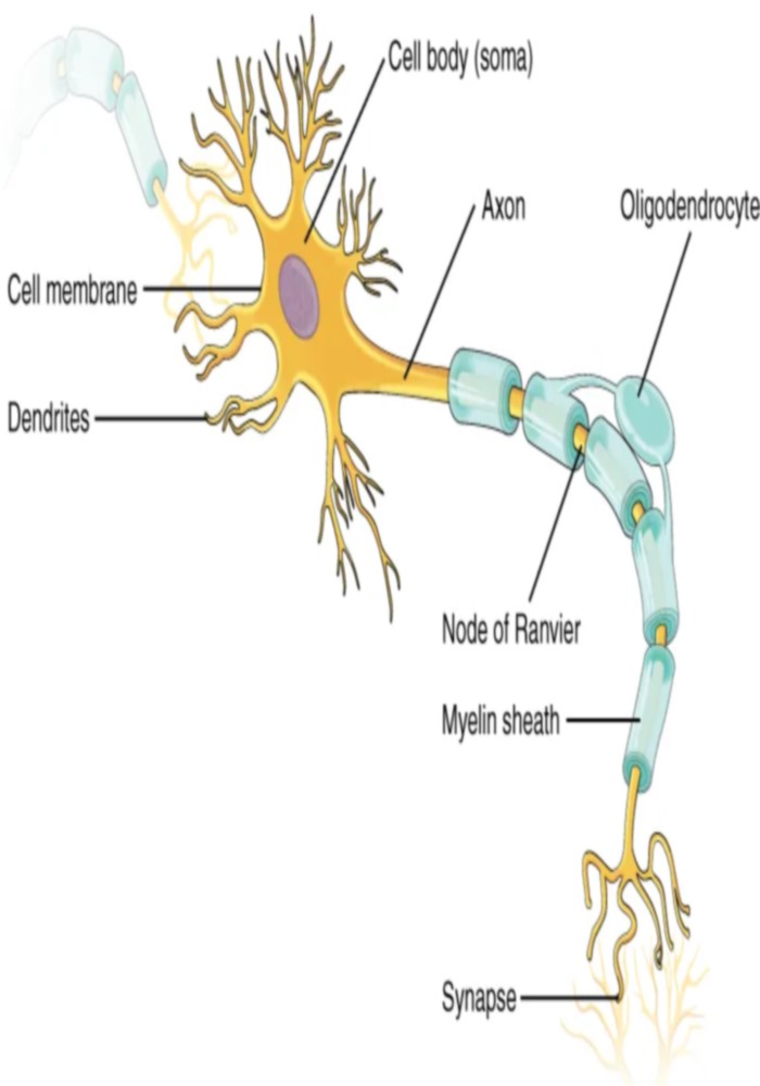
Nervous tissue is composed of two types of cells, neurons and glial cells. Neurons are the primary type of cell that most anyone associates with the nervous system. They are responsible for the computation and communication that the nervous system provides. They are electrically active and release chemical signals to target cells. Glial cells, or glia, are known to play a supporting role for nervous tissue. Ongoing research pursues an expanded role that glial cells might play in signaling, but neurons are still considered the basis of this function. Neurons are important, but without glial support they would not be able to perform their function.Neurons are the cells considered to be the basis of nervous tissue. They are responsible for the electrical signals that communicate information about sensations, and that produce movements in response to those stimuli, along with inducing thought processes within the brain. An important part of the function of neurons is in their structure, or shape...
Muscle Structure
Inside each skeletal muscle, muscle fibers are organized into individual bundles, each called a fascicle, by a middle layer of connective tissue called the perimysium. This fascicular organization is common in muscles of the limbs; it allows the nervous system to trigger a specific movement of a muscle by activating a subset of muscle fibers within a bundle, or fascicle of the muscle. Inside each fascicle, each muscle fiber is encased in a thin connective tissue layer of collagen and reticular fibers called the endomysium. The endomysium contains the extracellular fluid and nutrients to support the muscle fiber. These nutrients are supplied via blood to the muscle tissue. In skeletal muscles that work with tendons to pull on bones, the collagen in the three tissue layers (the mysia) intertwines with the collagen of a tendon. At the other end of the tendon, it fuses with the periosteum coating the bone.
Bone Structure
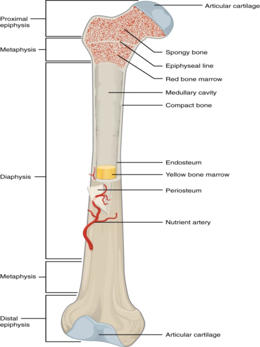
The structure of a long bone allows for the best visualization of all of the parts of a bone (Figure 6.7). A long bone has two parts: the diaphysis and the epiphysis. The diaphysis is the tubular shaft that runs between the proximal and distal ends of the bone. The hollow region in the diaphysis is called the medullary cavity, which is filled with yellow marrow. The walls of the diaphysis are composed of dense and hard compact bone The wider section at each end of the bone is called the epiphysis (plural = epiphyses), which is filled with spongy bone. Red marrow fills the spaces in the spongy bone. Each epiphysis meets the diaphysis at the metaphysis, the narrow area that contains the epiphyseal plate (growth plate), a layer of hyaline (transparent) cartilage in a growing bone. When the bone stops growing in early adulthood (approximately 18–21 years), the cartilage is replaced by osseous tissue and the epiphyseal plate becomes an epiphyseal line...
مدرسه پزشکی در مسیر موفقیت فرزندتان

فرم درخواست رزرو مصاحبه
شروع کلاس های جدید مدرسه پزشکی در مرداد ماه سال 1405
تمرکز اصلی بر روزهای چهارشنبه، پنجشنبه و جمعه
حضوری تهران و همزمان آنلاین در سراسر کشور
36 ماه آموزش تخصصی
پشتیبانی هر روزه در طول این 36 ماه آموزش تخصصی